
Ultrasound Podcast
Jailyn Avila
0
Ultrasound Podcast is a medical education show focused on cutting-edge bedside ultrasound and fresh takes on foundational ultrasound topics. It is presented as the original Ultrasound podcast and is aimed at clinicians who want practical, current information for point-of-care ultrasound. The show also directs listeners toward a year-long virtual ultrasound fellowship, free website content, courses, and a question bank. Its language and feed indicate a U.S. audience.
Эпизоды
-
 The Other Side of the Heart - The Neglected Ventrcle 29.06.2026 21минIn this episode, we discuss a video published on our YouTube channel on right heart ultrasound. We discuss advanced methods to differentiate acute versus chronic right heart enlargement, with emphasis on pulmonary embolism evaluation and risk stratification. They cover when right heart findings are most useful, including known PE and peri-arrest patients too unstable for CT. They review key views for RV size, favoring the apical four-chamber view, with discussion of parasternal and subcostal alternatives and limitations. For RV systolic function, they explain TAPSE and S' (abnormal if TAPSE
 -
Arthrocentesis in the Emergency Department 16.06.2026 25минIn this episode, Ben and Jailyn discuss how they approach arthrotencesis in ED patients. Watch the Youtube channel for a full run through for how to perform taps on the shoulder, knee, hip, elbow and ankle. 🌐 Connect with Us 🌐 If you want more education goodness, check out the links below! Check out our courses here: https://courses.coreultrasound.com Check out our question bank here: https://courses.coreultrasound.com/collections/q-bank Check out our FREE content on our website: https://www.coreultrasound.com Check out our in-person course in San Diego, Ca on Nov 12-14 2026 here: https://www.soundandsurf.com/essentials-2026 Disclaimer: By watching this (or any Core Ultrasound) education video or reading any of our content, you agree not to use this information as medical advice to treat any medical condition in either yourself or others, including but not limited to patients that you are treating. Consult your own physician for any medical issues that you may be having. This entire disclaimer applies to any and all content produced or discussed by Core Ultrasound. Under no circumstances shall any of the contributors on this website be responsible for damages arising from use of any topic discussed. This content should not be used in any legal capacity whatsoever, including but not limited to establishing "standard of care" in a legal sense or as a basis for expert witness testimony. No guarantee is given regarding the accuracy of any statements or opinions made by Core Ultrasound. The content of this website is formed by our own opinions and do not represent the views or opinions of any institutions we may be affiliated with.
-
How to Save a Life with POCUS (with Dr. Sophia Fornbacher, MD) 02.06.2026 16минIn this episode, we're thrilled to be joined by Dr. Sophia Fornbacher, ultrasound faculty and assistant professor at Riverside Community Hospital. Recently completing her ultrasound fellowship, Dr. Fornbacher shares her experiences and expertise on two remarkable cases where ultrasound made a critical difference. Spoiler alerts: Cardiac tamponade due to thoracic aortic dissections can trick you (without ultrasound at the bedside), and sometimes, 30cc/kg boluses aren't the best thing to do in patients with diabetic ketoacidosis (or hyperosmolar hyperketotic states)!
-
The RUSH Exam - Undifferentiated Shock POCUS 27.05.2026 17минWhen a patient rolls in hypotensive and tachycardic and you don't know why, the RUSH exam may be the most important tool in your hands. RUSH, Rapid Ultrasound in Shock and Hypotension, is a rapid, systematic bedside ultrasound protocol designed for exactly that moment. The HIMAP mnemonic breaks it down: (H)eart: The highest-yield component. Look for pericardial effusion/tamponade, right heart strain (think PE), or low EF. (I)VC: Not just a volume status tool, it reflects CVP and helps differentiate obstructive, distributive, and hypovolemic shock. Put a shocked septic patient on pressors and watch the IVC fill up even before you've given a drop of fluid. (M)orrison's Pouch aka the FAST Exam: Free fluid in the abdomen isn't just for trauma. Ruptured ectopic pregnancy, splenic rupture, ruptured bowel, and AAA all show up here. (A)orta: Rare but critical. Ruptured AAA and dissection can present as undifferentiated shock, and dissection matters more than people think, especially in patients with stroke-like symptoms plus pain. (P)ulmonary: Rule out tension pneumothorax At the bedside, ultrasound is the fastest and most accurate tool available for undifferentiated shock. Learn HIMAP. Use it early. 🎧 Happy scanning. 🌐 Connect with Us 🌐 If you want more education goodness, check out the links below! Check out our courses here: https://courses.coreultrasound.com Check out our question bank here: https://courses.coreultrasound.com/collections/q-bank Check out our FREE content on our website: https://www.coreultrasound.com Check out our in-person course in San Diego, Ca on Nov 12-14 2026 here: https://www.soundandsurf.com/essentials-2026 Disclaimer: By watching this (or any Core Ultrasound) education video or reading any of our content, you agree not to use this information as medical advice to treat any medical condition in either yourself or others, including but not limited to patients that you are treating. Consult your own physician for any medical issues that you may be having. This entire disclaimer applies to any and all content produced or discussed by Core Ultrasound. Under no circumstances shall any of the contributors on this website be responsible for damages arising from use of any topic discussed. This content should not be used in any legal capacity whatsoever, including but not limited to establishing "standard of care" in a legal sense or as a basis for expert witness testimony. No guarantee is given regarding the accuracy of any statements or opinions made by Core Ultrasound. The content of this website is formed by our own opinions and do not represent the views or opinions of any institutions we may be affiliated with.
-
Pericardial Effusion and When to Worry about Tamponade 19.05.2026 16минJailyn Avila and Ben Smith discuss a recent Core Ultrasound YouTube lecture on diagnosing pericardial effusions and cardiac tamponade with point-of-care ultrasound, emphasizing its importance in emergency medicine and cardiac arrest. The Reason trial (PMID: 27693280; Romolo Gasparin et al) demonstrates improved survival to ROSC when pericardial effusion is identified with ultrasound—a finding that contrasts sharply with Beck's triad, which shows poor sensitivity. Emergency department–based studies reveal that all three Beck's triad signs are rarely present simultaneously. The RUSH/HIMAP approach offers a structured assessment framework. Blind pericardiocentesis in PEA without prior ultrasound confirmation should be avoided. Key tamponade findings include: a dilated plethoric IVC with minimal collapse, RV diastolic collapse (including RVOT assessment and M-mode timing with valve opening), right atrial diastolic collapse, and ultrasound assessment of pulsus paradoxus—a finding more useful for ruling out tamponade than confirming it.
-
Ultrasound-Guided Thoracentesis 14.05.2026 11минJailyn and Ben kick off a new podcast format where they discuss the full-length lectures published on the Core Ultrasound YouTube channel — adding commentary, clinical pearls, and behind-the-scenes discussion that doesn't make it into the edited videos. Why this new podcast format? The YouTube videos are kept concise, but a lot of valuable clinical discussion gets left on the cutting room floor. This podcast fills that gap. In this episode, they break down their approaches to ultrasound-guided thoracentesis, with a detour into related tips for paracentesis. Static vs. Dynamic approach Static: Identify the pocket, mark the spot, proceed with a landmark technique Dynamic: Watch the needle in real-time as it enters the fluid When to use each: Large effusions → static often sufficient; small or complex effusions → dynamic preferred Identifying the right pocket Find the largest fluid pocket that is cephalad enough to clear the diaphragm and caudal enough to avoid pneumothorax Watch diaphragm excursion with respiration to confirm safe needle trajectory Avoid inadvertent liver/spleen biopsy Probe and needle technique for thoracentesis Use a phased array or curvilinear probe to find the effusion, then switch to linear for the procedure Orient transducer in the intercostal space (sagittal, rotated) In-plane (long axis) approach recommended — short axis is mechanically difficult due to the ribs Probe orientation: oblique when lateral, nearly horizontal when posterior Paracentesis pearls Out-of-plane technique can work well, given the larger pockets typical in the ED Curvilinear probe makes needle identification harder vs. linear For small-volume paras (e.g., ruling out SBP), dynamic approach is strongly preferred Color Doppler before you needle Scan the intended trajectory with color Doppler to rule out intercostal arteries or abdominal wall vessels (e.g., inferior epigastrics, caput medusa) Release probe pressure nearly completely when looking for veins — they collapse easily and can be missed Resources Mentioned 🎥 Full lecture video: Core Ultrasound YouTube Channel 📚 Premium courses: courses.coreultrasound.com Thanks for listening — and happy scanning!
-
 Measuring Cardiac Output Using Point-of-Care Ultrasound 26.01.2026 6минHow to Obtain Cardiac Output (5 Minute Sono Series) In this video, we'll guide you through the steps to calculate the cardiac output using the stroke volume and the patients heart rate. Learn how to capture VTI images with an A5C view and PW Doppler, calculate heart rate from the E-wave peaks, and combine stroke volume to determine cardiac output. Ideal VTI measurements and best practices for achieving accurate results are also covered. Join us for this informative session and happy scanning! 🌐 Connect with Us 🌐 If you want more education goodness, check out the links below! Check out our courses here: https://courses.coreultrasound.com Check out our question bank here: https://courses.coreultrasound.com/collections/q-bank Check out our FREE content on our website: https://www.coreultrasound.com Check out our longitudinal, year-long, and virtual ultrasound fellowship here: https://academy.ultrasoundleadershipacademy.com Disclaimer: By watching this (or any Core Ultrasound) education video or reading any of our content, you agree not to use this information as medical advice to treat any medical condition in either yourself or others, including but not limited to patients that you are treating. Consult your own physician for any medical issues that you may be having. This entire disclaimer applies to any and all content produced or discussed by Core Ultrasound. Under no circumstances shall any of the contributors on this website be responsible for damages arising from use of any topic discussed. This content should not be used in any legal capacity whatsoever, including but not limited to establishing "standard of care" in a legal sense or as a basis for expert witness testimony. No guarantee is given regarding the accuracy of any statements or opinions made by Core Ultrasound. The content of this website is formed by our own opinions and do not represent the views or opinions of any institutions we may be affiliated with.
 -
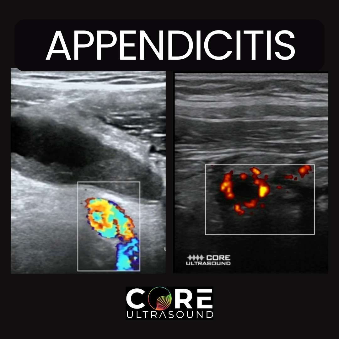 Appendicitis Diagnosis with Ultrasound 14.01.2026 8минIn this video, Jailyn explores the techniques and tips for diagnosing appendicitis using ultrasound. Learn about the preferred probes, patient preparation, and step-by-step guidance on locating the appendix. She covers common landmarks, such as the iliac artery, and important diagnostic criteria, including measurements and signs of inflammation. Real case examples demonstrate normal and abnormal findings, including appendicitis and perforated appendicitis. This comprehensive guide is essential for healthcare professionals aiming to enhance their diagnostic skills in ultrasound imaging. 🌐 Connect with Us 🌐 If you want more education goodness, check out the links below! Check out our courses here: https://courses.coreultrasound.com Check out our question bank here: https://courses.coreultrasound.com/com Check out our FREE content on our website: https://www.coreultrasound.com Disclaimer: By watching this (or any Core Ultrasound) education video or reading any of our content, you agree not to use this information as medical advice to treat any medical condition in either yourself or others, including but not limited to patients that you are treating. Consult your own physician for any medical issues that you may be having. This entire disclaimer applies to any and all content produced or discussed by Core Ultrasound. Under no circumstances shall any of the contributors on this website be responsible for damages arising from use of any topic discussed. This content should not be used in any legal capacity whatsoever, including but not limited to establishing "standard of care" in a legal sense or as a basis for expert witness testimony. No guarantee is given regarding the accuracy of any statements or opinions made by Core Ultrasound. The content of this website is formed by our own opinions and do not represent the views or opinions of any institutions we may be affiliated with.
 -
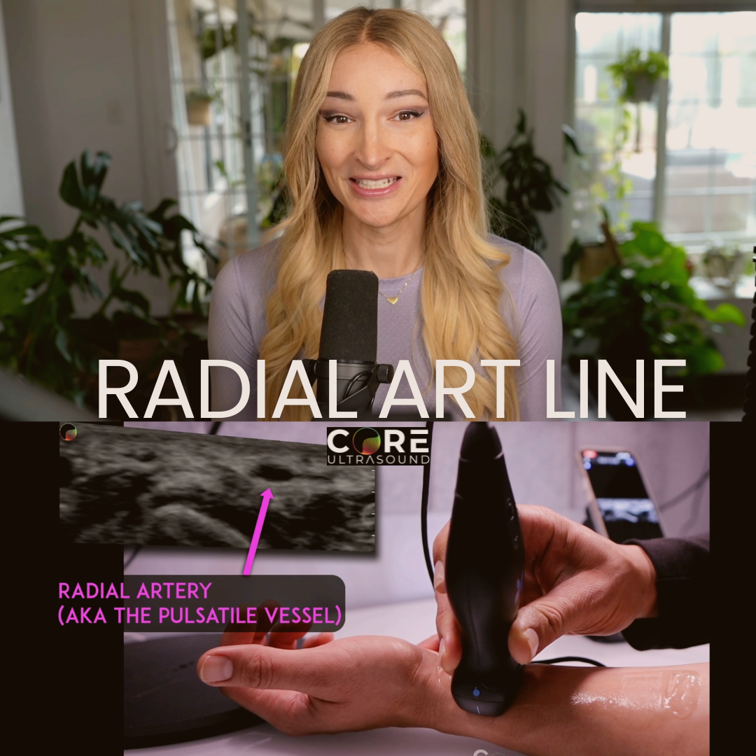 How to Place Arterial Catheters Using POCUS 05.01.2026 6минoin Jailyn Avila as she provides a comprehensive guide on placing a radial arterial line using ultrasound guidance. This five-minute(ish) tutorial covers the necessary equipment, including the linear transducer and radial artery catheterization set, as well as the sterilization protocols required for the procedure. Jailyn walks through each step of needle insertion, sequential needle tip tracking, guidewire placement, and catheter advancement. She also shares valuable tips on identifying blood vessels, ensuring accurate catheter positioning, and securing the line. Additionally, Jailyn highlights important considerations such as patient prep and the use of local anesthetics to enhance patient comfort. 🌐 Connect with Us 🌐 If you want more education goodness, check out the links below! Check out our courses here: https://courses.coreultrasound.com Check out our question bank here: https://courses.coreultrasound.com/collections/q-bank Check out our FREE content on our website: https://www.coreultrasound.com Check out our longitudinal, year-long, and virtual ultrasound fellowship here: https://academy.ultrasoundleadershipacademy.com Disclaimer: By watching this (or any Core Ultrasound) education video or reading any of our content, you agree not to use this information as medical advice to treat any medical condition in either yourself or others, including but not limited to patients that you are treating. Consult your own physician for any medical issues that you may be having. This entire disclaimer applies to any and all content produced or discussed by Core Ultrasound. Under no circumstances shall any of the contributors on this website be responsible for damages arising from use of any topic discussed. This content should not be used in any legal capacity whatsoever, including but not limited to establishing "standard of care" in a legal sense or as a basis for expert witness testimony. No guarantee is given regarding the accuracy of any statements or opinions made by Core Ultrasound. The content of this website is formed by our own opinions and do not represent the views or opinions of any institutions we may be affiliated with.
 -
 Aortic Stenosis: Identification and Quantification Made Simple 01.12.2025 13минIn this week's video, we delve into how to diagnose aortic stenosis using Point of Care Ultrasound at the bedside. After watching this video, you'll know how to identify and quantify aortic stenosis, understand its severity, and recognize why it's considered the deadliest valvular disease. Discover techniques ranging from basic visual inspections to advanced methods like the continuity equation, all tailored for bedside clinicians. Plus, get insights on using ultrasound for effective diagnosis. Ideal for emergency room settings and beyond. Happy scanning! By watching this (or any Core Ultrasound) education video or reading any of our content, you agree not to use this information as medical advice to treat any medical condition in either yourself or others, including but not limited to patients that you are treating. Consult your own physician for any medical issues that you may be having. This entire disclaimer applies to any and all content produced or discussed by Core Ultrasound. Under no circumstances shall any of the contributors on this website be responsible for damages arising from use of any topic discussed. This content should not be used in any legal capacity whatsoever, including but not limited to establishing "standard of care" in a legal sense or as a basis for expert witness testimony. No guarantee is given regarding the accuracy of any statements or opinions made by Core Ultrasound. The content of this website is formed by our own opinions and do not represent the views or opinions of the any institutions we may be affiliated with.
 -
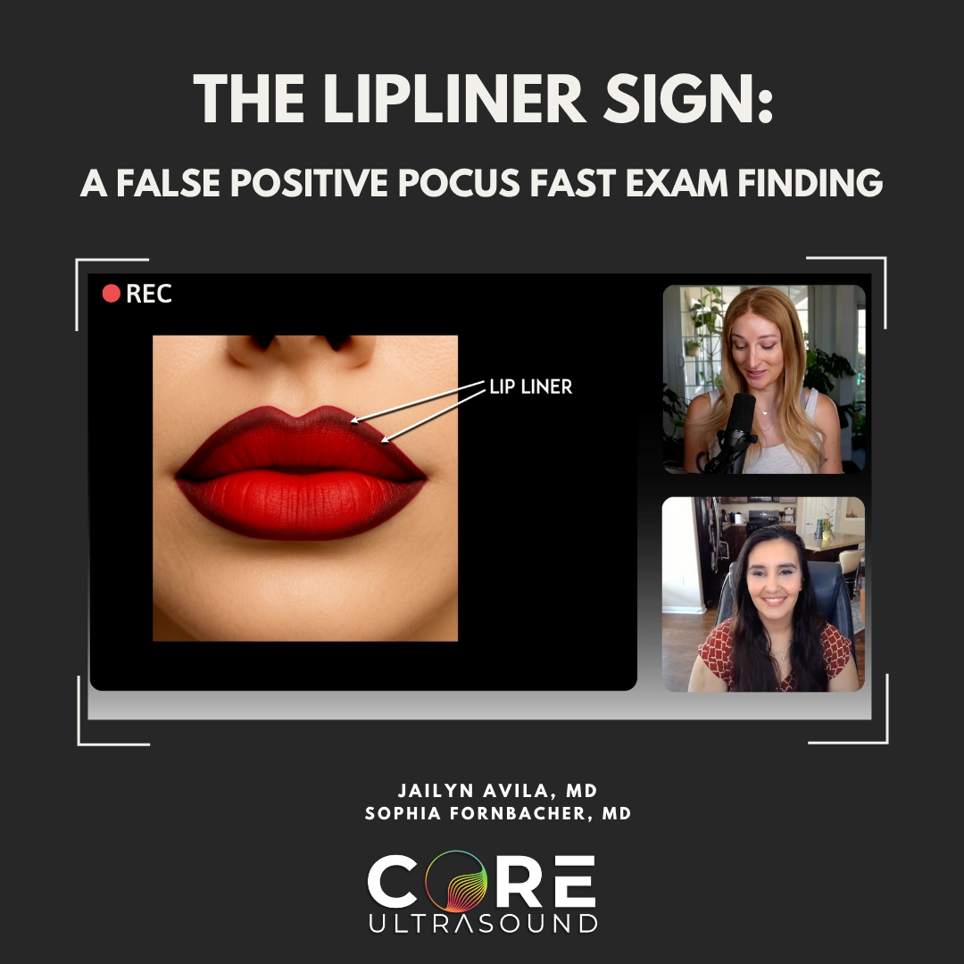 Exploring the Lip Liner Sign: Avoiding False Positives in FAST Examinations 18.11.2025 18минExploring the Lip Liner Sign: Avoiding False Positives in FAST Examinations with Dr. Sophia Fornbacher In this episode, host Jailyn Avila sits down with Dr. Sophia Fornbacher, a fellowship-trained emergency medicine physician, to discuss the intricacies of the lip liner sign—a recently published artifact that can lead to false positives in FAST (Focused Assessment with Sonography for Trauma) examinations. They delve into their experiences and thoughts on the FAST exam's relevance in various clinical settings, and the importance of recognizing artifacts like the lip liner sign to improve diagnostic accuracy. Additionally, they touch on other related signs and nomenclatures, while also reflecting on their teaching experiences and their approach to ultrasound education. 🌐 Connect with Us 🌐 If you want more education goodness, check out the links below! Check out our longitudinal, year-long and virtual ultrasound fellowship here: https://academy.ultrasoundleadershipacademy.com Check out our FREE content on our website: https://www.coreultrasound.com Check out our courses here: https://courses.coreultrasound.com Check out our question bank here: https://courses.coreultrasound.com/collections/q-bank Disclaimer: By watching this (or any Core Ultrasound) education video or reading any of our content, you agree not to use this information as medical advice to treat any medical condition in either yourself or others, including but not limited to patients that you are treating. Consult your own physician for any medical issues that you may be having. This entire disclaimer applies to any and all content produced or discussed by Core Ultrasound. Under no circumstances shall any of the contributors on this website be responsible for damages arising from use of any topic discussed. This content should not be used in any legal capacity whatsoever, including but not limited to establishing "standard of care" in a legal sense or as a basis for expert witness testimony. No guarantee is given regarding the accuracy of any statements or opinions made by Core Ultrasound. The content of this website is formed by our own opinions and do not represent the views or opinions of the any institutions we may be affiliated with.
 -
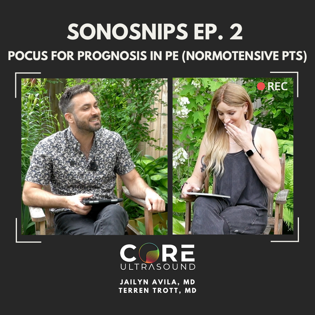 Sonosnips (ICU POCUS) Episode 2! (US in Normotensive PE) 11.11.2025 13минIn this episode of the Core Ultrasound Podcast, Dr. Jailyn Avila and Dr. Terren Trott discuss the prognostic value of echocardiography in normotensive patients with acute pulmonary embolism (PMID: 24412192). We analyze a 2014 study published in the Journal of American College of Cardiology, which involved 411 patients who were initially normotensive. The study found that parameters like TAPSE less than 15 millimeters and the RV to LV ratio in the apical four-chamber view had the best predictive value for adverse outcomes. This episode offers insights into how these findings can be applied in clinical practice to improve patient management.
 -
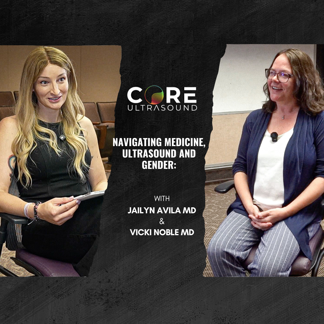 Navigating Medicine, Ultrasound and Gender: An Inspiring Interview with Dr. Vicki Noble 09.06.2025 34минJoin me this week as I share an inspiring interview with Dr. Vicki Noble, Chair of Emergency Medicine at University Hospitals Cleveland Medical Center. We discuss her illustrious career, her role as a leader in point-of-care ultrasound, and her experiences navigating the medical field as a woman. Dr. Noble also shares valuable insights on the importance of collaboration, the future of medical training, and advice for women pursuing careers in medicine. This episode highlights the significance of community and mentorship in the field of emergency medicine. Don't forget to check out the Women in Medicine Conference in Chicago from September 18-20, 2025. For more information and to register, visit: WIMedicine.org. Enjoy the interview and happy scanning! 🌐 Connect with Us 🌐 If you want more educational goodness, check out the links below! Check out our FREE content on our website: https://www.coreultrasound.com Check out our courses here: https://courses.coreultrasound.com Check out our question bank here: https://courses.coreultrasound.com/collections/q-bank Disclaimer: By watching this (or any Core Ultrasound) education video or reading any of our content, you agree not to use this information as medical advice to treat any medical condition in either yourself or others, including but not limited to patients that you are treating. Consult your own physician for any medical issues that you may be having. This entire disclaimer applies to any and all content produced or discussed by Core Ultrasound. Under no circumstances shall any of the contributors on this website be responsible for damages arising from use of any topic discussed. This content should not be used in any legal capacity whatsoever, including but not limited to establishing "standard of care" in a legal sense or as a basis for expert witness testimony. No guarantee is given regarding the accuracy of any statements or opinions made by Core Ultrasound. The content of this website is formed by our own opinions and do not represent the views or opinions of the any institutions we may be affiliated with.
 -
 Artificial Intelligence and POCUS: Insights from the Experts! (Part 2) 29.05.2025 20минExploring AI and Distance Learning in Ultrasound Education with Drs Michael Prats and Ben Smith. In this week's episode of the Core Ultrasound Podcast, we continue our in-depth exploration of innovative teaching methods for ultrasound education. Joined by Ben Smith, a full professor and interim chair at the University of Tennessee at Chattanooga, and Michael Prats, an Associate Professor and Director of Ultrasound Research at Ohio State University, explore the role of artificial intelligence (AI) in medical education, particularly in bedside ultrasound. We discuss the potential benefits, limitations, and future implications of AI in enhancing teaching and learning experiences for both educators and residents. Tune in to gain insights on the evolving landscape of ultrasound education and the promising intersection of AI in this field. 🌐 Connect with Us 🌐 If you want more educational goodness, check out the links below! Check out our longitudinal, year-long and virtual ultrasound fellowship here: https://academy.ultrasoundleadershipacademy.com Check out our FREE content on our website: https://www.coreultrasound.com Check out our courses here: https://courses.coreultrasound.com Check out our question bank here: https://courses.coreultrasound.com/collections/q-bank Disclaimer: By watching this (or any Core Ultrasound) education video or reading any of our content, you agree not to use this information as medical advice to treat any medical condition in either yourself or others, including but not limited to patients that you are treating. Consult your own physician for any medical issues that you may be having. This entire disclaimer applies to any and all content produced or discussed by Core Ultrasound. Under no circumstances shall any of the contributors on this website be responsible for damages arising from use of any topic discussed. This content should not be used in any legal capacity whatsoever, including but not limited to establishing "standard of care" in a legal sense or as a basis for expert witness testimony. No guarantee is given regarding the accuracy of any statements or opinions made by Core Ultrasound. The content of this website is formed by our own opinions and do not represent the views or opinions of the any institutions we may be affiliated with.
 -
 "Ultrasound Without Experts? How AI and Tele-Guidance Are Stepping In (Part 1, with Mike Prats!) 20.05.2025 27минIn this week's episode, join host Jailyn Avila and special guest Dr. Michael Prats, an Associate Professor and Director of Ultrasound Research at Ohio State University, as they delve into the dynamic world of ultrasound. Discover advancements in ultrasound technology, its applications in pre-hospital settings, and the potential of AI in medical training. From engaging discussions on birdwatching to practical insights into patient self-performed ultrasounds, this episode covers a broad spectrum of topics that aim to enhance healthcare accessibility and effectiveness. Tune in for an enlightening conversation that bridges practical experience with emerging technologies, transforming the landscape of healthcare education. Stay tuned for Part 2, where we add in Ben Smith to the conversation! 🌐 Connect with Us 🌐 If you want more education goodness, check out the links below! Check out our longitudinal, year-long and virtual ultrasound fellowship here: https://academy.ultrasoundleadershipacademy.com Check out our FREE content on our website: https://www.coreultrasound.com Check out our courses here: https://courses.coreultrasound.com Check out our question bank here: https://courses.coreultrasound.com/collections/q-bank Disclaimer: By watching this (or any Core Ultrasound) education video or reading any of our content, you agree not to use this information as medical advice to treat any medical condition in either yourself or others, including but not limited to patients that you are treating. Consult your own physician for any medical issues that you may be having. This entire disclaimer applies to any and all content produced or discussed by Core Ultrasound. Under no circumstances shall any of the contributors on this website be responsible for damages arising from use of any topic discussed. This content should not be used in any legal capacity whatsoever, including but not limited to establishing "standard of care" in a legal sense or as a basis for expert witness testimony. No guarantee is given regarding the accuracy of any statements or opinions made by Core Ultrasound. The content of this website is formed by our own opinions and do not represent the views or opinions of the any institutions we may be affiliated with.
 -
Thoracic Aortic Emergencies on Ultrasound: Recognizing Aneurysms and Dissections 01.05.2025 25минIn this video, Dr. Jailyn Avila walks you through how to diagnose thoracic aortic aneurysms and thoracic aortic dissection using bedside ultrasound. While the abdominal aorta often gets the most attention, aortic disease above the diaphragm is just as critical—and often harder to recognize. Early detection of thoracic aortic pathology can be life-saving, and ultrasound can play a key role in raising suspicion quickly. Timestamps: 0:58 - Intro 3:25 - Basics of scanning 6:35 - Thoracic aortic aneurysm 11:12 - Thoracic aortic dissection 🌐 Connect with Us 🌐 Check out our longitudinal, year-long and virtual ultrasound fellowship here: https://ultrasoundleadershipacademy.com Check out our FREE content on our website: https://www.coreultrasound.com Check out our courses here: https://courses.coreultrasound.com Check out our question bank here: https://courses.coreultrasound.com/collections/q-bank Disclaimer: By watching this (or any Core Ultrasound) education video or reading any of our content, you agree not to use this information as medical advice to treat any medical condition in either yourself or others, including but not limited to patients that you are treating. Consult your own physician for any medical issues that you may be having. This entire disclaimer applies to any and all content produced or discussed by Core Ultrasound. Under no circumstances shall any of the contributors on this website be responsible for damages arising from use of any topic discussed. This content should not be used in any legal capacity whatsoever, including but not limited to establishing "standard of care" in a legal sense or as a basis for expert witness testimony. No guarantee is given regarding the accuracy of any statements or opinions made by Core Ultrasound. The content of this website is formed by our own opinions and do not represent the views or opinions of any institutions we may be affiliated with.
-
Ultrasound-Guided Interfascial Plane Block for Shoulder Pain | ED Applications with Dr. Gabe Rose 16.04.2025 27минIn this episode, we sit down with Dr. Gabe Rose (@kpsono, co-author of a new study published in the American Journal of Emergency Medicine, to explore the ultrasound-guided Interfascial Plane Block (IPB) — a promising technique for treating acute shoulder pain in the emergency department. ✅ We cover: What the Interfascial Plane Block is and how it works Key anatomical landmarks and ultrasound guidance Advantages over the traditional interscalene block for shoulder pain Safety considerations and ideal patient selection 📄 Read the study: "Treatment of shoulder pain with ultrasound-guided interfascial plane block" https://doi.org/10.1016/j.ajem.2024.10.022 🎥 Want more videos on ultrasound-guided regional anesthesia and POCUS education? Subscribe to us, and send us your questions and experiences with shoulder nerve blocks! 🔗 Follow Dr. Gabe Rose on social media: Instagram: https://www.instagram.com/doc.gabe/ TikTok: https://www.tiktok.com/@doc.gabe Follow KP Sono: Instagram: https://www.instagram.com/kp.sono/ TikTok: https://www.tiktok.com/@kp.sono 🌐 Connect with Us 🌐 Check out our longitudinal, year-long and virtual ultrasound fellowship here: https://ultrasoundleadershipacademy.com Check out our FREE content on our website: https://www.coreultrasound.com Check out our courses here: https://courses.coreultrasound.com Check out our question bank here: https://courses.coreultrasound.com/collections/q-bank Disclaimer: By watching this (or any Core Ultrasound) education video or reading any of our content, you agree not to use this information as medical advice to treat any medical condition in either yourself or others, including but not limited to patients that you are treating. Consult your own physician for any medical issues that you may be having. This entire disclaimer applies to any and all content produced or discussed by Core Ultrasound. Under no circumstances shall any of the contributors on this website be responsible for damages arising from use of any topic discussed. This content should not be used in any legal capacity whatsoever, including but not limited to establishing "standard of care" in a legal sense or as a basis for expert witness testimony. No guarantee is given regarding the accuracy of any statements or opinions made by Core Ultrasound. The content of this website is formed by our own opinions and do not represent the views or opinions of any institutions we may be affiliated with.
-
Game-Changer for Shoulder Pain: The Interfascial Plane Block 02.04.2025 4минIn this video, we demonstrate the technique for performing an ultrasound-guided interfascial plane block (IPB)—a simple, effective, and motor-sparing alternative to the interscalene block for treating shoulder pain in the Emergency Department. This block targets the axillary nerve within the plane between the deltoid and subscapularis muscles and has shown promise in providing significant pain relief while avoiding diaphragmatic paralysis and upper extremity motor dysfunction. 🩺 Special thanks to Drs. Billy Shank, Tyler Yates, and Gabe Rose for their contributions to this video and for helping bring this important technique to life. 🔗 Follow Dr. Gabe Rose on social media: Instagram: https://www.instagram.com/doc.gabe/ TikTok: https://www.tiktok.com/@doc.gabe Follow KP Sono: Instagram: https://www.instagram.com/kp.sono/ TikTok: https://www.tiktok.com/@kp.sono 🌐 Connect with Us 🌐 Check out our longitudinal, year-long and virtual ultrasound fellowship here: https://ultrasoundleadershipacademy.com Check out our FREE content on our website: https://www.coreultrasound.com Check out our courses here: https://courses.coreultrasound.com Check out our question bank here: https://courses.coreultrasound.com/collections/q-bank Disclaimer: By watching this (or any Core Ultrasound) education video or reading any of our content, you agree not to use this information as medical advice to treat any medical condition in either yourself or others, including but not limited to patients that you are treating. Consult your own physician for any medical issues that you may be having. This entire disclaimer applies to any and all content produced or discussed by Core Ultrasound. Under no circumstances shall any of the contributors on this website be responsible for damages arising from use of any topic discussed. This content should not be used in any legal capacity whatsoever, including but not limited to establishing "standard of care" in a legal sense or as a basis for expert witness testimony. No guarantee is given regarding the accuracy of any statements or opinions made by Core Ultrasound. The content of this website is formed by our own opinions and do not represent the views or opinions of any institutions we may be affiliated with.
-
Ultrasound-Guided Carpal Tunnel Release: A New ED Solution? 18.03.2025 27минCarpal tunnel syndrome is a common condition seen in the emergency department, but traditional treatment options often involve long wait times for definitive care. Could ultrasound-guided carpal tunnel release (CTR) be a game-changer for ED management? In this interview, Dr. Gabe Rose discusses how this minimally invasive technique can be used in the ED, its benefits over traditional approaches, and what emergency physicians need to know to implement it. We cover: ✅ The basics of ultrasound-guided CTR✅ Advantages over conventional surgical release✅ Indications and patient selection in the ED ✅ Technical considerations for emergency physicians Click here to learn how to diagnose CTS! Follow Dr. Gabe Rose: Instagram: https://www.instagram.com/doc.gabe/ TikTok: https://www.tiktok.com/@doc.gabe Follow KP Sono: Instagram: https://www.instagram.com/kp.sono/ TikTok: https://www.tiktok.com/@kp.sono 🌐 Connect with Us 🌐 Check out our longitudinal, year-long and virtual ultrasound fellowship here: https://ultrasoundleadershipacademy.com Check out our FREE content on our website: https://www.coreultrasound.com Check out our courses here: https://courses.coreultrasound.com Check out our question bank here: https://courses.coreultrasound.com/collections/q-bank Disclaimer: By watching this (or any Core Ultrasound) education video or reading any of our content, you agree not to use this information as medical advice to treat any medical condition in either yourself or others, including but not limited to patients that you are treating. Consult your own physician for any medical issues that you may be having. This entire disclaimer applies to any and all content produced or discussed by Core Ultrasound. Under no circumstances shall any of the contributors on this website be responsible for damages arising from use of any topic discussed. This content should not be used in any legal capacity whatsoever, including but not limited to establishing "standard of care" in a legal sense or as a basis for expert witness testimony. No guarantee is given regarding the accuracy of any statements or opinions made by Core Ultrasound. The content of this website is formed by our own opinions and do not represent the views or opinions of any institutions we may be affiliated with.
-
How Ultrasound Can Transform Cardiac Arrest Resuscitation (Part 2!) 30.12.2024 14минIn part two of this series on using ultrasound during cardiac arrest, I dive into advanced strategies to further optimize your resuscitation care: 1️⃣ Avoiding Pulse Check Delays: Learn how to use ultrasound efficiently during pulse checks to minimize interruptions in chest compressions and maintain high-quality CPR. 2️⃣ Resuscitation Modifications: Discover practical modifications and techniques that integrate ultrasound into your resuscitation workflow, improving team performance and patient outcomes. Building on the foundation from part one, this video focuses on actionable tips to take your ultrasound skills to the next level during cardiac arrest scenarios. 🔗 If you haven't seen part one yet, make sure to check it out for the essentials of using ultrasound to identify reversible causes, guide procedures, and support resuscitation! 🌐 Connect with Us 🌐 Check out our longitudinal, year-long and virtual ultrasound fellowship here: https://ultrasoundleadershipacademy.com Check out our FREE content on our website: https://www.coreultrasound.com Check out our courses here: https://courses.coreultrasound.com Check out our question bank here: https://courses.coreultrasound.com/collections/q-bank Disclaimer: By watching this (or any Core Ultrasound) education video or reading any of our content, you agree not to use this information as medical advice to treat any medical condition in either yourself or others, including but not limited to patients that you are treating. Consult your own physician for any medical issues that you may be having. This entire disclaimer applies to any and all content produced or discussed by Core Ultrasound. Under no circumstances shall any of the contributors on this website be responsible for damages arising from use of any topic discussed. This content should not be used in any legal capacity whatsoever, including but not limited to establishing "standard of care" in a legal sense or as a basis for expert witness testimony. No guarantee is given regarding the accuracy of any statements or opinions made by Core Ultrasound. The content of this website is formed by our own opinions and do not represent the views or opinions of any institutions we may be affiliated with.
Похожие подкасты


Популярен в
Этот подкаст также попадал в подкаст-чарты этих стран.
